Docket #: S18-383
Theranostic Nanoparticles for Identification and Reduction of Myeloid Cell-Mediated Immunosuppression in Cancer
Stanford researchers in the Richard Zare Lab have developed dual-function nanoparticles that selectively accumulate in immune-suppressing myeloid cells in tumors, allowing these cells to be both imaged and treated with a single agent.
Myeloid-derived suppressor cells (MDSCs) and related myeloid cells build up in many cancers, where they shield tumors from the immune system and reduce the effectiveness of treatment. Current drugs that alter myeloid cell behavior also affect other immune and non-immune cells, causing harmful side effects. Blood tests can count these cells but cannot track their movement into tumors.
The Stanford nanoparticles are coated with granulocyte-colony stimulating factor (G-CSF), a signaling protein that regulates myeloid cells and steers the particles into them. Each particle can carry an imaging dye, a therapeutic drug, or both. In a mouse model of triple-negative breast cancer, coated particles accumulated in MDSCs within the tumor and spleen at significantly higher rates than uncoated particles. A single dose of particles carrying an anti-inflammatory drug shrank tumors by about 25%, while tumors in the control group grew more than 20% within three days.
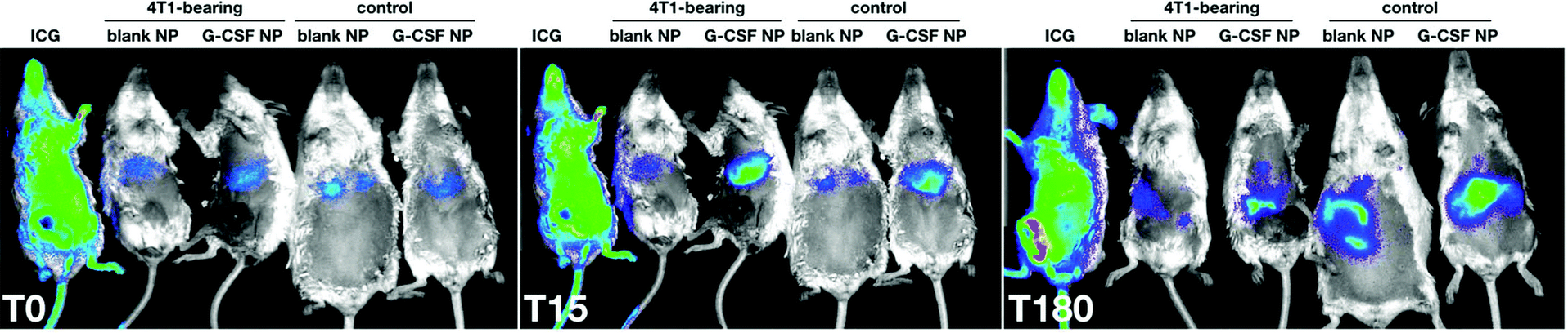
Image Description: In vivo imaging of blank and G-CSF-decorated nanoparticles in 4T1 breast carcinoma-bearing and control mice at 0, 15, and 180 minutes after injection. (Image courtesy Margulis et al. (2020) Nanoscale)
Stage of Development
Applications
- Targeted therapy to reduce myeloid-driven immunosuppression in solid tumors such as breast, lung, colorectal and prostate cancer, melanoma, and glioma
- In vivo imaging of myeloid cell location and tumor infiltration for prognosis, patient stratification, and treatment monitoring
Advantages
- All components have prior FDA approval
- No off-target side effects: Nanoparticles preferentially accumulate in myeloid cells
- > 80% retention of drug payload within 5 hours of exposure to biological medium
- Imaging and therapy in one particle
- Selective: G-CSF coating significantly increases uptake by MDSCs, with limited nonspecific uptake (under 10% of T cells)
- Stable payload: keeps more than 80% of its cargo within 5 hours in biological media, without an early "burst" release
- Faster path to the clinic: built entirely from FDA-approved components
- Single-dose tumor shrinkage in a preclinical model
Publications
- Margulis et al. Nanoscale (2020) "Nanoparticles Decorated with Granulocyte-Colony Stimulating Factor for Targeting Myeloid Cells"
Related Links
Patents
- Published Application: WO2021127000
- Published Application: 20220370644
Similar Technologies
-
Surface-Enhanced Raman Scattering Nanoparticles for Imaging Tumor Oxidative Stress S19-246Surface-Enhanced Raman Scattering Nanoparticles for Imaging Tumor Oxidative Stress
-
Oligocarbonate molecular transporters across biological barriers S08-441Oligocarbonate molecular transporters across biological barriers
-
Phase-change nanoparticles for neuromodulation S17-163Phase-change nanoparticles for neuromodulation